Cartilage Mapping
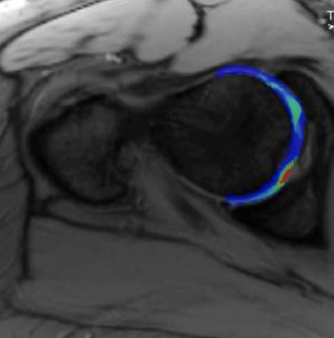
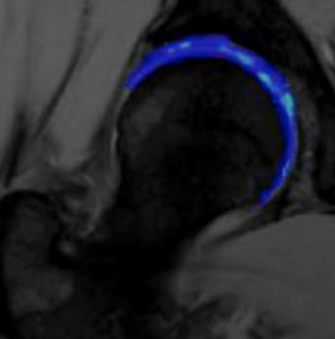
Cartilage mapping scanning technology has advanced to a point where we can not only see the amount of cartilage that is present in the joint, we can also get an idea of the quality and health of it! There are a few ways of doing this, the original being termed dGEMRIC (delayed Gadolinium Enhanced MRI of Cartilage). This requires an intravenous injection of contrast (Gadolinium). The patient then walks around for 20-45 minutes and then has their scan. It is very time consuming and the intravenous injection can be an issue if you have kidney problems. Therefore, other techniques have been developed; the best one (in our view!) is T2 cartilage mapping. This does not require injections of any sort, although it does add approximately 5 minutes to your scan!
Although all of these techniques are still termed ‘experimental’ the information from the cartilage mapping helps us determine the health of your joint and the areas that may be more damaged than others. This helps the surgeon give you a more accurate prognosis about what may or may not be achieved by various surgical options.


RECOGNISED BY ALL THE MAJOR INSURERS


