CT & Motion Analysis
Many different factors, and often combinations of pathologies, can cause hip pain. Our job is to unpick these so that we can help get you on the road to recovery. These, as mentioned throughout our website, rely on accurate diagnosis. Most sports injuries related to the hip joint such as labral tears are secondary to mild developmental abnormalities such as femoroacetabular impingement (FAI) and/or subtle dysplasia (shallow hip socket). These can be difficult to diagnose with plain x-rays and even MRI scans.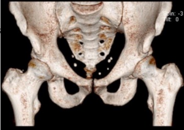
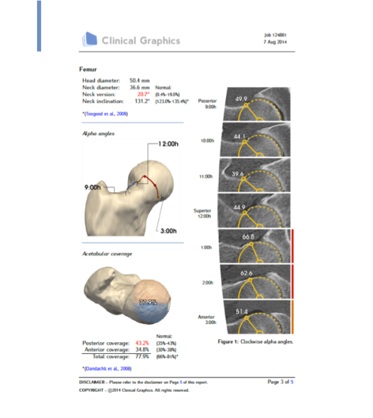
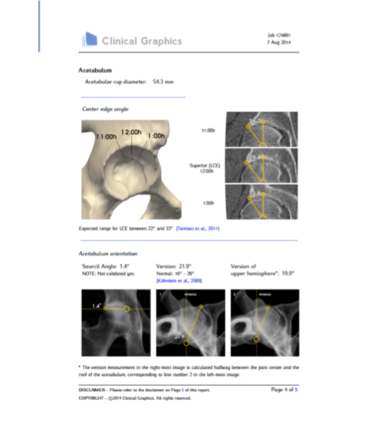
Currently, the best way to look at the shape of the bones is CT (computerised tomography) scans, from which you can get 3D reconstructions. This is like looking at your skeleton and is a fantastic way of assessing the way the actual joint is formed.
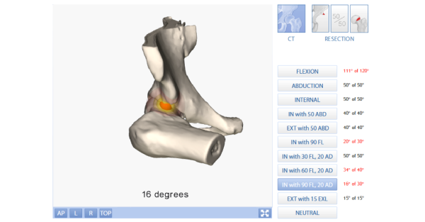
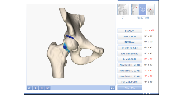
However, things have moved on even further. From the CT scans we can see the way your hip actually moves by sending the scans off for ‘Motion Analysis’. We work with Stryker’s HipMap team, who specialise in breaking the scans down and rebuilding them showing us how your hip moves. This can demonstrate areas of impingement or dysplasia graphically, both helping you understand your own body, but also demonstrating where the bones should be reshaped to ensure accurate surgery and the best results.
There is a possible downside though. Like x-ray, CT does expose the patient to radiation. However, the current dose from our protocol is estimated to be the equivalent of three x-rays, and so is very low.
RECOGNISED BY ALL THE MAJOR INSURERS


