MRI
Accurate diagnosis relies on obtaining information and piecing it together to obtain  what is usually a ‘best guess’. In medicine, this will always be the case to a greater or lesser extent as everyone is different, and not all symptoms of the same disease present in the same way. It is surprising how often people with a clear case of arthritis can be treated for months for something completely different such as muscle strains
or back pain.
Therefore we like to get the most information about you as we can, and make sure that what we get is as accurate as it can be. Most MRI scanners, for instance, are termed 1.5 Tesla. This refers to the strength of the magnet that the scanner has. The magnet in the scanner is used to align water in your body, which ‘spins’ as the magnet is turned on and off (as water is a charged particle). As the water particles spin, they give of a signal, which is what the scanner detects. Depending on where that water is and what it is part of, determines how quickly it will spin and therefore affects the signal the water gives off. It’s very complicated! However, in simple terms, the stronger the magnet, the more spin, and therefore the stronger signal. This leads to clearer scans, which helps give a more accurate diagnosis.
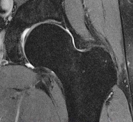
Historically, to diagnose a labral tear accurately in a 1.5T scanner, you would need a doctor to inject your hip joint with a large volume dye prior to the scan (termed an arthrogram). This is because the clarity of the scans from a 1.5T scanner tends to be poor (around the hip) and the dye gives a better contrast. The arthrogram is performed under local anaesthetic, but the vast majority of patients who have had this done do not wish for another, as it can be quite uncomfortable. However, some people still consider this technique the ‘gold standard’.
Even so, MRI arthrograms have been reported to have a few problems. One is that they can ‘over-call’ labral tears (as in suggest there is one present when there is not). The other is that generally they tend to be very dark scans, which means that the doctor cannot look at the other muscles and structures around the joint which can also be implicated in your pain, it purely looks at the labrum.
With the introduction of 3 Tesla MRI scanners, the arthrogram injection is no longer needed as the clarity of the scans is so much better. This means a faster visit to the hospital, no injection or pain, and in fact more information as all the surrounding hip structures can be assessed as well.
RECOGNISED BY ALL THE MAJOR INSURERS


